Bo Yin1,
Jian-Jun Ji1 ![]() ,
Ming Yang2
,
Ming Yang2
For correspondence:- Jian-Jun Ji Email: jianjun066@hotmail.com Tel:+865375250877
Received: 27 October 2015 Accepted: 6 August 2016 Published: 30 September 2016
Citation: Yin B, Ji J, Yang M. Polymeric implant of methylprednisolone for spinal injury: preparation and characterization. Trop J Pharm Res 2016; 15(9):1833-1837 doi: 10.4314/tjpr.v15i9.3
© 2016 The authors.
This is an Open Access article that uses a funding model which does not charge readers or their institutions for access and distributed under the terms of the Creative Commons Attribution License (http://creativecommons.org/licenses/by/4.0) and the Budapest Open Access Initiative (http://www.budapestopenaccessinitiative.org/read), which permit unrestricted use, distribution, and reproduction in any medium, provided the original work is properly credited..
Purpose: To improve the effectiveness and reduce the systemic side effects of methylprednisolone in traumatic spinal injuries, its polymeric implants were prepared using chitosan and sodium alginate as the biocompatible polymers.
Methods: Implants of methylprednisolone sodium succinate (MPSS) were prepared by molding the drug-loaded polymeric mass obtained after ionotropic gelation method. The prepared implants were evaluated for drug loading, in vitro drug release and in vivo performance in traumatic spinal-injury rat model with paraplegia.
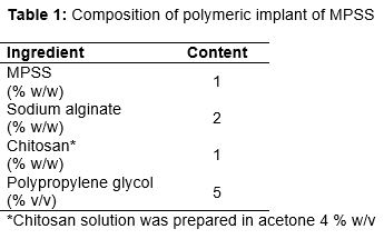
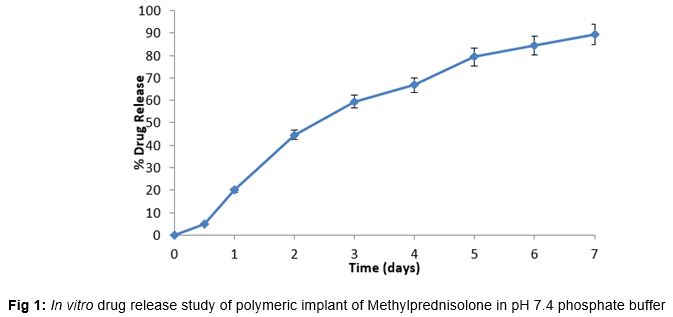
Results: All the implant formulations were light pale solid matrix with smooth texture. Implants showed 86.56 ± 2.07 % drug loading. Drug release was 89.29 ± 1.25 % at the end of 7 days. Motor function was evaluated in traumatic spinal injury-induced rats in terms of its movement on the horizontal bar. At the end of 7 days, the test group showed the activity score (4.75 ± 0.02) slightly higher than that of standard (4.62 ± 0.25), but the difference was not statistically different (p > 0.05).
Conclusion: MPSS-loaded implants produces good recovery in traumatic spinal-injury rats.
Introduction
Implants are invasive polymeric delivery devices which are intended to release the drug in the injected/inserted body cavity in a very slow manner for prolonged periods of time which may extend from a week to few months and even to many years. These may be prepared either from non-biodegradable polymers or from biodegradable polymers. Non-biodegradable implants are needed to be removed by another surgical procedure after the expected completion of drug in the polymeric implant. Though invasive nature of procedures is a limitation of use of implant, easy termination of therapy (if required in case of adverse effects) by removal of implant is a unique advantage of implants. Unlike conventional parenteral dosage forms drug delivery by implants can reduce dosage frequency, provide prolonged duration of action and improve the patient compliance [1,2]. Implants can deliver drug in the microenvironment of target site or in vicinity of target site with maximizing the drug utilization and providing local as well as systemic effects. Implants may be solid matrix type or reservoir type devices with or without special drug releasing mechanisms like molds, tablets, pellets, osmotic pumps, infusion pumps etc. or may be in situ gelling systems which upon injection get converted in to a gel with contact of body fluids, change in temperature or pH [3,4].
Spinal column is the most sophisticated and complex biological target site for drug delivery. Traumatic spinal cord injury is one of the most complex injuries to manage. Spinal injury leads to permanent disability, paraplegia, tetraplegia and over all decreased life expectancy [5-7]. There is no cure of spinal cord injury but efforts can be made to cope with the effects of primary and secondary spinal injury.
The management of neuropathic pain and inflammation is the prime objective in treatment of spinal injuries. Many potent nonsteroidal anti-inflammatory drugs (NSAIDs) and steroids (in high dosage) are administered for the same. But to provide the prolonged and target specific drug delivery various NSAIDs and steroids have been investigated for intrathecal administration so as to provide improved therapeutic effect [8,9]. Various biodegradable polymers have been investigated for preparing scaffolds or polymeric network/platform/implant of developing for neuroprotective and neuroregenerative drug delivery systems in spinal cord injuries [10,11].
Methylprednisolone sodium succinate (MPSS) is the glucocorticoid used in effective management of traumatic spinal injury [12]. It has been reported to show significant recovery from tissue damage, neuropathic pain and inflammation. Studies have also shown improvement in motor nerve activity with the use of MPSS in spinal injury [13,14]. This study aims to develop the polymeric implantable device of MPSS and to study its effect on traumatic spinal injured rats with paraplegia.
Methods
Materials
Methylprednisolone sodium succinate (MPSS), chitosan 75 % deacylated and sodium alginate (SA) were obtained from Sigma Aldrich Japan. Rests of the chemicals were of analytical grade.
Preparation of polymeric hydrogels of MPSS
Implants of MPSS were prepared by molding of drug loaded polymeric mass obtained after the ionotropic gelation method (). Accurately weighed quantity of sodium alginate was dispersed in distilled water and stirred at 200 rpm for 30 min meanwhile weighed quantity of the drug was dispersed onto the dispersion of alginate. This drug-alginate solution was mixed with 4 % solution of chitosan in acetone with stirring with addition of propylene glycol. Then this solution was kept in teflon molds with flat bottom and slowly 2 % aqueous calcium chloride solution was sprayed for 10 minutes till the mass attained the gel like consistency. The molds containing polymeric matrix loaded with the drug was frozen at -10 ± 1 oC for rigidization overnight. The prepared implant molds were taken out to room temperature and then cut into small cubic pieces of desired size with a surgical blade. For hardening these implants were kept in a desiccator rich in vapors of formaldehyde which were produced by keeping 37 % v/v formaldehyde solution at the bottom of the desiccator for 48 h. After hardening, the implants were taken out, air dried for 4 days for removing traces of formaldehyde from the implants. Finally the prepared implants were sterilized by ethylene oxide and then packed in butter paper for storage in cool dark place in an aseptic environment.
Drug loading
One implant (15 × 15 × 15 mm) was dissolved in 50 ml of methanol with continuous stirring (200 rpm) at 37 ± 1 oC) for 1 h (n=6). One mL sample was withdrawn, diluted suitably and then analysed spectrophotometrically.
In vitro release of drug
In vitro drug release study was performed according to a reported method [15]. One implant (15 × 15 × 15 mm) was kept in a tightly closed vial with pH 7.4 buffer (10 ml) as media. Several such vials were kept on an incubator shaker (at 37 ± 1.0 oC). For taking sample a single vial was withdrawn at definite time interval till 7 days and analysed spectrophotometrically for drug release (n = 3).
In vivo study on traumatic spinal-injury rats
Healthy male Wistar rats (200 - 240 g) were used for the study. After procurement, experiment was carried after giving 3 - 4 days of resting and acclimatization. The rats were fed standard diet and water ad libitum. Protocols of the study were duly approved (approval no. 2014/A243) by Animal Ethical Committee of Peking University. In vivo study was executed as per directives of European Commission on animal handling [16].
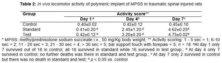
A total of 54 rats were divided in three groups (control, standard and test group) of eighteen rats each. All the animals were subjected to laminectomy (after injecting thiopental sodium 40 mg/kg) by giving the acute spinal injury by compression method (extradural 50 g force clip for 10 s at around T2) laterally after giving thiopental sodium (40 mg/kg). Immediately the control groups were administered the non-medicated implant. The test group received the polymeric implant of MPSS (50 mg/Kg body weight; i.t.) near injury. MPSS was injected i.v. at 30 mg/Kg body weight in the Standard after 1 h of injury (and given the same at every alternate day for one week). At the end of 7 days, the locomotor activity was observed by the performance of rats in Horizontal Bar. The experiment was performed on an apparatus with a horizontal bar (2 mm diameter; 40 cm long) fitted on two support of 50 cm height. By holding the mouse tail it was left on the bar with the help of its forepaw’s contact in the center of the bar. The time spent by rats in horizontal bar was studied and scoring was done on the basis of following scale: 1-5 sec = 1; 6 - 10 sec = 2; 11 - 20 sec = 3; 21- 30 sec = 4; > 30 sec = 5; bar support touch with forepaw = 5.
Statistical analysis
The results are expressed as mean ± standard deviation (SD). P < 0.05 was considered statistically significant.
Results
Chitosan and alginate blend based polymeric implants loaded with MPSS were prepared by ionotropic gelation method and characterized for drug loading, in vitro drug release and in vivo performance in traumatic spinal injured rats. The method used is simple and easy to reproduce.
The implants were observed as the light pale solid matrix with smooth texture. Implants showed 86.56 ± 2.07 % drug loading. Drug release was 89.29 ± 1.25 % at the end of 7 days (). Alginate chitosan blend of polymer have been reported to provide controlled drug release in various previous studies [17,18].
Locomotor activity is governed by motor nerve activity which is adversely affected by spinal injury. Therefore, motor activity of the traumatic spinal injured rats were compared after administering the prepared implants of MPSS with that of standard (i.v. MPSS). Motor function was evaluated in terms of the rat’s movement in a horizontal bar (). After the 24 h no improvement in activity was observed in any group (activity score 0.40 ± 0.02). Moreover, out of 18 rats in control group only 7 survived. All 18 survived in standard (activity score: 0.41 ± 0.20) and 16 survived in test group (activity score: 0.42 ± 0.12) with insignificant improvement. As survival was quite good in test group animals it was inferred that the polymeric device was well tolerated in the subjects.
On the day 4, there was still no significant improvement in activity (activity score: 0.42 ± 0.12) in control group which remain only 4. The standard group showed activity score of 2.45 ± 1.20 at the end of day 4 with little tail lifting. On the other hand the test group showed better activity than that of standards with activity score of 3.20 ± 0.20.
At the end of the day 7 only 2 rats survived in control with no sign of improvement in the activity score (0.45 ± 0.10). The standard group showed an activity score of 4.62 ± 0.25 while the test group had an activity score of 4.75 ± 0.02 which is insignificantly (p < 0.05) different from that of standard.
Discussion
It has been shown by various previous studies that the implants prepared from biodegradable polymers are one of the most potential approaches of delivering drugs to target site. Delivering a drug in the microenvironment of target site ensures the maximum drug utilization with minimum drug dosage. Moreover, as drug is not distributed peripherally much, the adverse effects of the drug are also minimized [19,20]. In polymeric devices selection of biocompatible polymers and/or solvents is crucial to minimize chances of rejection of the devices by the subjects [21].
Biodegradable polymers have been investigated for various pharmaceutical drug delivery systems. Apart from being nontoxic, non-mutagenic and non-cytotoxic, the biodegradable polymers are metabolized in the body and eliminated by normal physiological pathways [22,23]. Methylprednisolone is a potent anti-inflammatory glucocorticoid which is used as a very first line treatment in cases of post traumatic SCI. It was reported to show significant recovery from primary tissue damage and secondary neuropathic pain and inflammation. Studies have also shown improvement in motor nerve activity with use of Mp in SCI [14,15,24].
Most of the studies have demonstrated positive effects of MPSS in SCI with parenteral (i.v.) administration. But high dosage of MPSS is also associated with the systemic adverse effects. Therefore, alternative routes or devices (like biodegradable implants) for administration of MPSS have been explored and investigated. Biodegradable implantable drug delivery techniques have been designed to release drugs at the targeted site like fractured vertebra, injured spinal cord etc. for prolonged period of time. But it requires surgical procedure for administering the implant devices to the target site [2-4].
In a recent study, in situ gel prepared from chitosan and PLGA (1:14) was found to be very potential in the post traumatic spinal injuries. The in situ gel showed high drug content, satisfactory rheological properties, prolonged drug release and the best in vivo anti-inflammatory and motor function activity performance [25]. This recent study also supported the use of implant for delivering MPSS in SCI. The prepared implant of MPSS showed the high % drug loading which is the prerequisite for the drug delivery systems for accommodating desired quantity of drug within the delivery matrix for subsequent slow diffusion or release of the drug. The in vitro drug release study of the prepared implant showed a slow and prolonged drug release which had been well supported by the various previous studies dealing with the alginate chitosan polymeric drug delivery matrix [17,18]. The prepared implants of MPSS showed motor activity in the traumatic spinal injured rats that was comparable to that of the standard (intravenous MPSS).
Conclusion
The findings of this study indicate that MPSS implants show high drug loading, prolonged in vitro drug release and good tolerance in rats. The implants also produces significant recovery in traumatic spinal injured rats. These implants may be useful for providing prolonged drug release and improved recovery in patients of traumatic spinal injury, but this has to be first confirmed in human experiments.
Declarations
Acknowledgement
References
Archives


News Updates